Initial Visualization
Aorta and IVC
- Good place to start ultrasound
- Teaches anatomic planes, identification of fluid
- Can be lifesaving – AAA
- IVC an help with fluid status, other
Narration
In this module we are going to talk about ultrasound visualization of the aorta and the inferior vena cava or IVC. This is a good place to start with ultrasound for a couple reasons. One, it gives you a sense of two of the primary planes we are going to be using in ultrasound- the transverse and sagittal plane, and also it is quite clinically useful. Visualization of a normal aorta or finding a AAA (abdominal aortic aneurysm) can actually be life-saving and visualization of the IVC can help with fluid status and other pathology.
Probe Selection & Positioning
- Curvilinear
- Between xiphoid process and umbilicus
- Gentle but firm pressure
- Start transverse


Narration
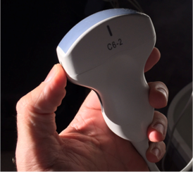
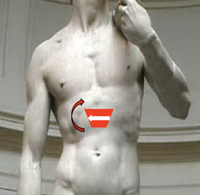
We will start with the curvilinear probe, I like to start in the transverse plane with the indicator to the patients right or our left as we are thinking of looking from the feet upwards just as we do in a ct scan. The key is to use gentle but firm pressure to move that bowel out of the way and you can see the entire aorta between the xyphoid process at the inferior border of the rib cage and the umbilicus and again we’ll start transverse and then move to sagittal.
Aorta & IVC
- Basic transverse view
- Aorta on vertebral body
- IVC to right


Narration
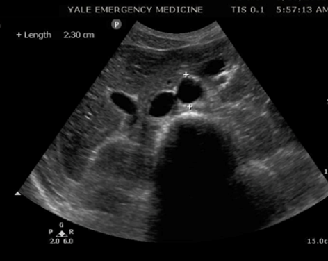
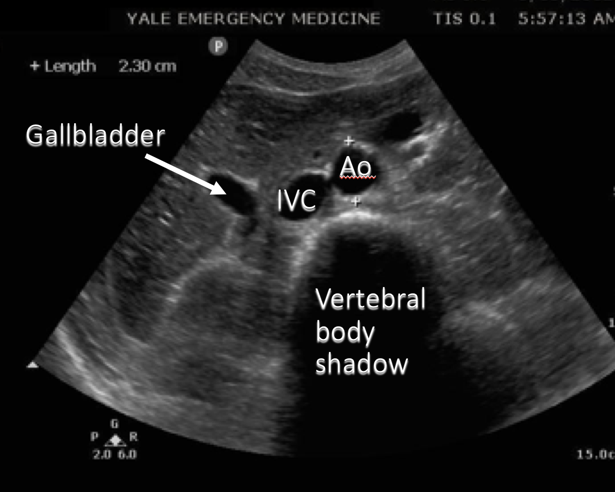
Here’s a labeled image that is a little bit larger, again we have the depth fairly deep about 15 cm, the indicator to the patients right and you can see that aorta which is measured in this case sitting right on top of the vertebral body to the patients left, the IVC is just to the right of that or left on the screen and you can see the gall bladder in this particular image.
Aorta/IVC Window

Narration
Here’s a labeled moving image, we’ve taken the depth up a little bit but we can still see the vertebral body shadow on a transverse view. That aorta is sitting right on top of the vertebral body and we have the IVC to the patients right with the left renal vein actually draining into the IVC.
Rotating Transverse To Sagittal

Narration
In this case what we are doing is starting in a transverse view, you can see the vertebral body in the far field initially you can see the aorta on the patients left and the IVC on the patients right, and then we are rotating this probe clockwise so the indicator is towards the patients head, keeping the aorta in view. If we are on the right side of the patient when we are scanning, this means the indicator is going to go towards our left or the patients head. This is a really good way to make sure you are visualizing the aorta and also to get a sense of how you are rotating from the transverse to the sagittal plane.
?
v:1 | onAr:0 | onPs:1 | tLb:1 | tLbJs:0uStat: False | db:0 | shouldInvoke:True | pu:False | em: | i: 0 | p: | pv:1 | refreshTime: 1/1/0001 12:00:00 AM | now: 2/19/2026 1:10:13 AM | c: