Transverse Aorta Proximal to Distal
Proximal Aorta

Narration
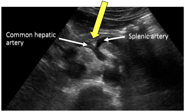
We’ll start by visualizing the abdominal aorta in the transverse plane with the indicator to the patients right or our left as we are looking from the feet upwards. We can usually visualize the entire abdominal aorta between the xyphoid process at the inferior edge of the rib cage and the umbilicus. Ideally, we can go all the way from the first branch of abdominal aorta which is the celiac artery, down to the bifurcation into the iliacs. In practice this can be challenging sometimes particularly in obese patients. You can be reasonably confident that if you see at least the midportion of the aorta and there is no aneurysm that the patient doesn’t have one, as this is typically where you will find it. So we are starting here with the proximal aorta and you can see that first branch which is the celiac and it is branching into the common hepatic artery going to the patient’s right and the splenic artery to the patient’s left.
Celiac Artery

"Seagull sign"
Narration
Here’s the schematic of that transverse view of that first branch, the celiac artery, sometimes call this the seagull sign, it looks like the wings of a seagull. You see the common hepatic artery to the right and the splenic artery to the left. There is also the left gastric branch of the celiac which you often don’t see that well on ultrasound.
Proximal To Mid Aorta
Narration
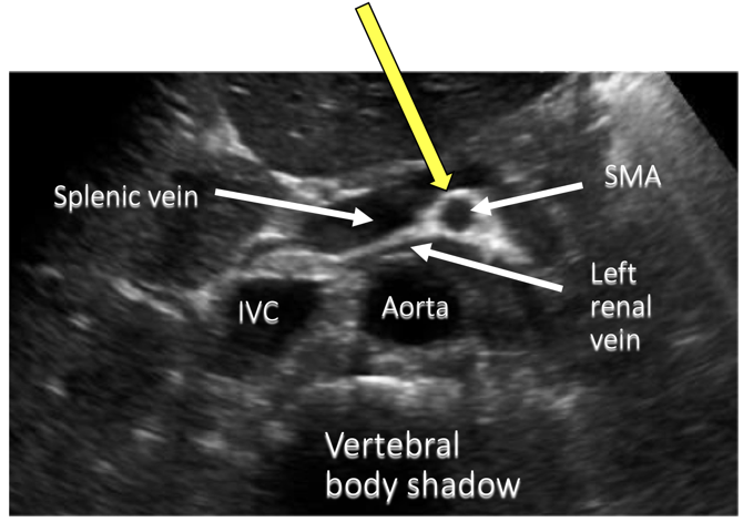
Continuing down a little bit, you can either slide or tilt the probe slightly, you can see the superior mesenteric artery in cross section. This is the second branch of the abdominal aorta. It is surrounded by some fatty tissue and we sometimes call this the mantle clock configuration.
Superior Mesnteric Artery

"Mantle clock"
Narration
Here’s a schematic and you can see that SMA that circular cross-sectional view right in the center with the aorta in the further part of the field sitting on the vertebral body. The splenic vein will cross anterior to the superior mesenteric artery and drain into the portal vein. You can see the IVC off to the patients right.
Mid-Aorta
Narration
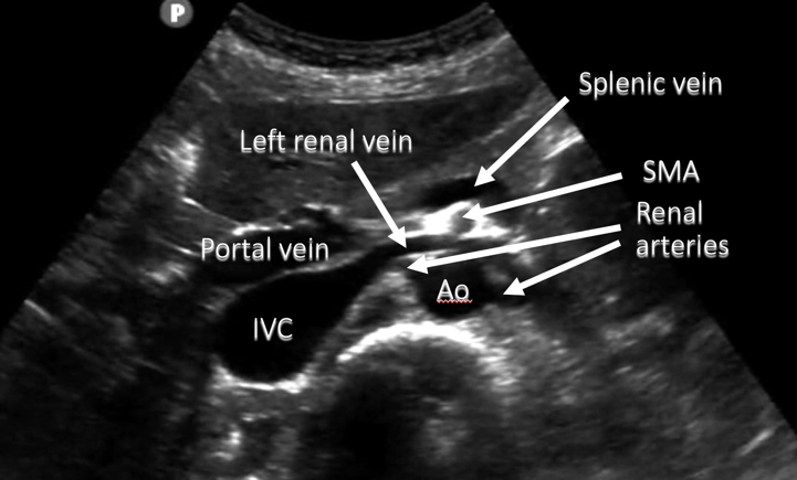
Here is another moving image of the mid aorta you can see the vertebral body in the far field, the aorta pulsing, and then sitting on top of that is the superior mesenteric artery with the left renal vein draining between the aorta and the SMA into the IVC and again you can see the splenic vein draining into the portal system which is anterior to the SMA.
Mid Aorta
Narration
Here’s a schematic showing this anatomy. Here’s the aorta sitting on top of the vertebral body with the right and left renal arteries coming out which really happens right about the level of the SMA and this is also typically where you’ll find most aneurysms. Also seen here is the left renal vein and the splenic vein.
Distal Aorta: Bifurcation Into Iliacs

Narration
As we go distally, we can either tilt the probe or slide it slightly we’ll see bifurcation into the iliac vessels at the distal part of the aorta.
This website is only for professional medical education. Contact your doctor for medical care. 2026 © MedEdu LLC. All Rights Reserved. Terms & Conditions | About Us | Privacy | Email Us